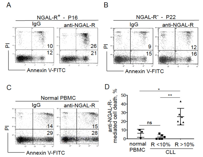
Figure 4.
Anti-NGAL-R antibodies induce the death in CLL cells. (A–C) Representative cytograms of (A) NGAL-R+ CLL cells (44%; P16), (B) NGAL-R− CLL cells (0%; P22) and (C) normal PBMCs cultured for 24 h in the presence or absence of 20 μg/mL anti-NGAL-R antibodies or rabbit IgG (isotype control); detection of apoptotic cells after annexin-V-FITC/PI staining and flow cytometry. Results are as log PI fluorescence intensity (y-axis) vs. log annexin-V-FITC fluorescence intensity (x-axis). The percentage of annexin-V-positive cells is shown. (D) Quantification of anti-NGAL-R-mediated survival levels in PBMCs from healthy individuals and CLL cells from untreated patients. The percentage of anti-NGAL-R-mediated survival is determined by subtracting the percentage of annexin-positive cells in the presence of rabbit IgG from the percentage of annexin-positive cells in the presence of anti-NGAL-R Ab. The data are presented as mean ± SD (normal PBMCs, n = 3; CLL cells, n = 12, with n = 6 NGAL-R+ (≥10%) and n = 6 NGAL-R− (<10%); Statistical relevance was assessed with the unpaired t-test; not significant (ns); * p < 0.05; ** p < 0.01.

