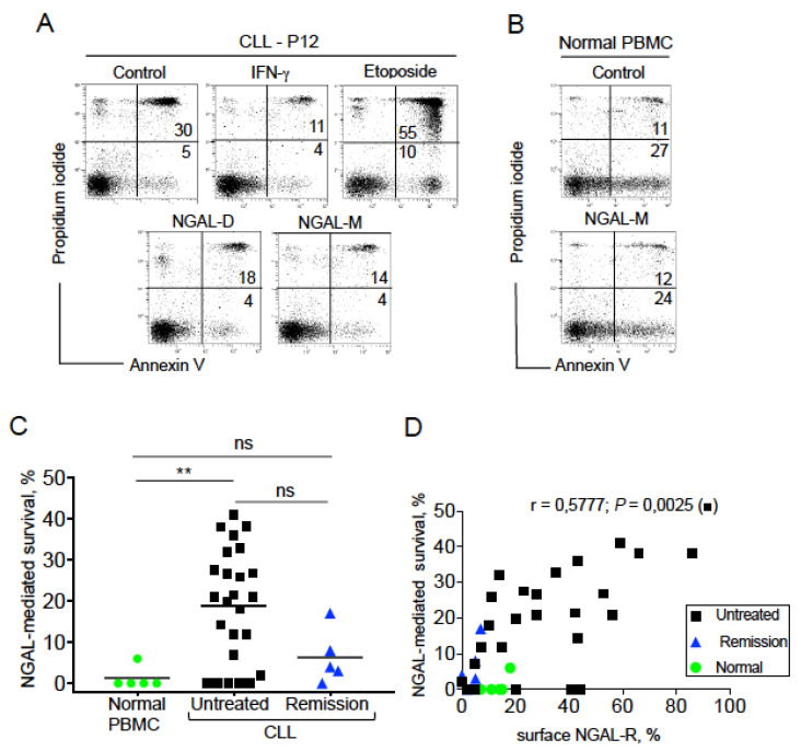
Figure 5.
NGAL protects CLL cells from spontaneous death. (A) Representative cytograms of CLL cells cultured for 24 h in the presence or absence of 100 nM recombinant human NGAL (dimers and monomers), 1000 U/mL IFN-γ or 1 μM etoposide; detection of apoptotic cells after annexin-V-FITC/PI staining and flow cytometry. The percentage of annexin-V-positive cells is shown. (B) Representative cytograms of PBMCs from one healthy donor cultured for 24 h in the presence or absence of 100 nM NGAL monomers, and cell death was assessed as described in (A). (C) NGAL-mediated survival levels were determined in PBMCs from healthy individuals and CLL cells from patients before treatment or at remission. The percentage of NGAL-mediated survival is determined by subtracting the percentage of annexin-positive cells in the absence of NGAL from the percentage of annexin-positive cells in the presence of NGAL, and divided by the percentage of annexin-positive cells in the absence of stimulus x 100. The data are presented as mean ± SEM (normal PBMCs, n = 5; CLL cells from untreated patients, n = 25; CLL cells from patients in remission, n = 5). p values were calculated using a Mann–Whitney U-test; not significant (ns); ** p < 0.01. (D) Correlation between the levels of surface NGAL-R and NGAL-mediated survival in normal PBMCs (n = 5), CLL cells from untreated patients (n = 25) and PBMCs from patients in remission (n = 5); Spearman’s correlation coefficient (r) and the p-value are shown for untreated CLL.

