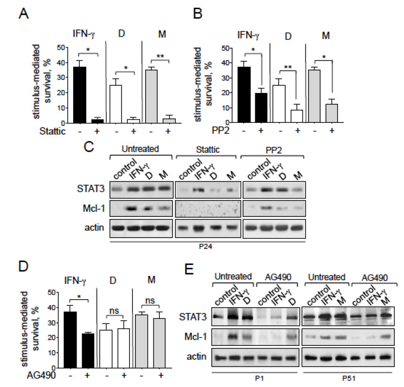
Figure 7.
Exogenous NGAL induces CLL cell survival through the Src/STAT3/Mcl-1 signaling pathways. (A–C) CLL cells were cultured for 24 h in the presence or absence of NGAL dimers or monomers (100 nM) or IFN-γ (1000 U/mL) after a 15 min pretreatment with (A) 5 μM STAT3 inhibitor, (B) 10 μM PP2 (an Src family kinase inhibitor), or (D) 10 μM AG490 (a JAK2 inhibitor); after which NGAL-mediated survival was assessed as described in Figure 4. The data are presented as mean ± SEM (n = 3). Statistical relevance was assessed with the paired t-test; not significant (ns); * p < 0.05; ** p < 0.01. (C,E) CLL cells were cultured for 24 h in the presence or absence of NGAL dimers or monomers (100 nM) or IFN-γ (1000 U/mL) after a 15 min pretreatment with (C) 5μM STAT3 inhibitor, 10 μM PP2, or (E) 10 μM AG490; after which lysates were Western blotted (reducing conditions) with antibodies against STAT3, Mcl-1 and actin. Representative experiments (n = 3) are shown.

