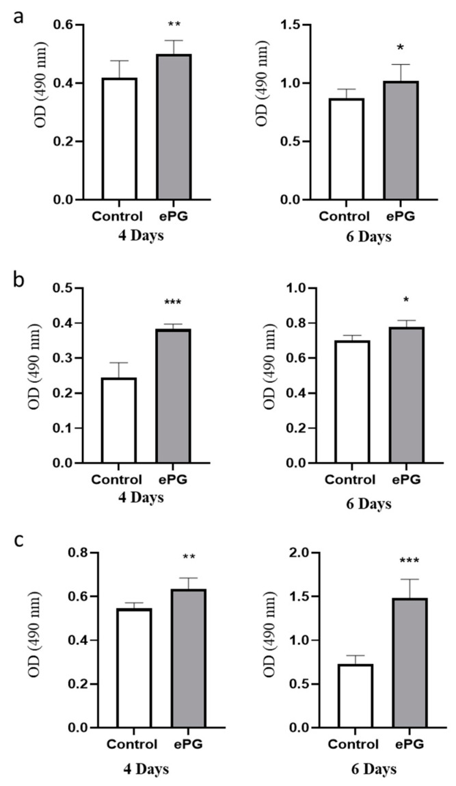
Figure 5.
Enhanced proliferation induced by ePG in three human PDAC cell lines. (a) PANC1 cells, (b) MIA PaCa-2, and (c) BxPC-3 cells were cultured in hypoxia alone or with ePG (MOI 10). Extracellular bacteria were killed with antibiotics after one hour of infection, and cell proliferation was determined by MTS after 4 or 6 days. Two-tailed t-test was performed for statistical analysis. * p ≤ 0.05, ** p ≤ 0.01, *** p ≤ 0.005.

