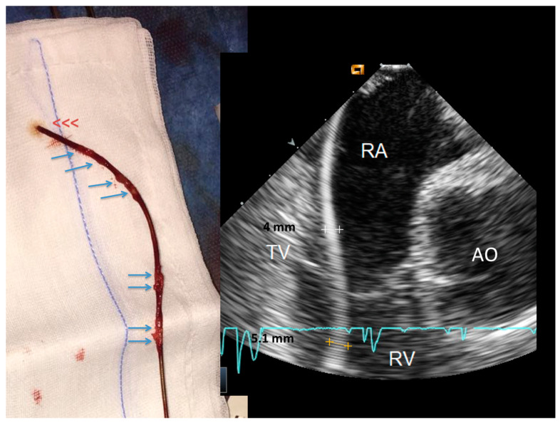
Figure 2.
Side-by-side, a thickened lead at ICE and the same lead at visual inspection immediately after extraction. The ventricular lead (right side) is uniformly thickened at ICE. After extraction, the lead (left side) shows remnants of sheathing fibrosis in both the ventricular and atrial parts of the lead (arrows). Abbreviations as in Figure 1.

