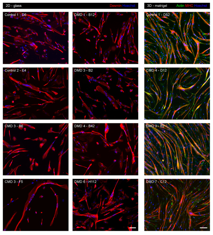
Figure 3.
Myogenic differentiation of iHMuSC clones. Clones selected for their good growth capacity were tested for their myogenic differentiation capacity. In the 2D glass condition (left and middle panels), clones were differentiated on glass culture supports for 5 days before the detection of desmin (red) using immunofluorescence. In the 3D-Matrigel condition (right panel), clones were differentiated between 2 thin Matrigel coats for 5 days before the detection of actin (green) and MHC (red) using immunofluorescence. Nuclei are labelled with Hoechst (blue). Bars = 100 µm.

