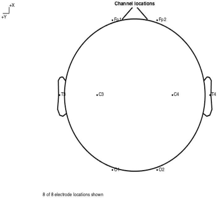
Figure A1.
Schematic of electrode locations. All recordings were performed using an eight-electrode montage, with bilateral temporal (Fp1 and Fp2), temporal (T3 and T4), parietal (C3 and C4) and occipital (O1 and O2) electrodes, positioned per the 10–20 system. All recordings were carried out under resting state conditions. To minimise behavioural disturbance, patients were allowed to spend time adjusting to the recording environment and recordings were carried out for extended sessions, with the first several minutes discarded.

