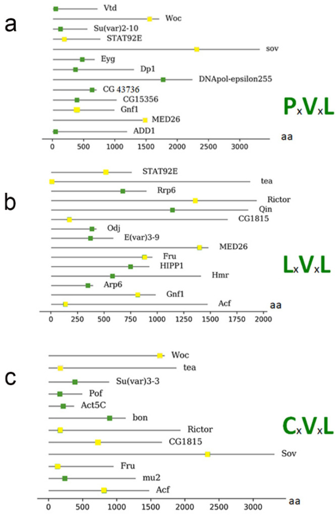
Figure 3.
Representation of proteins that have a possible motif for interaction with HP1a from Table 2. (a) The proteins connected to the motif PxVxL and the location of the motif within the amino acid sequence. (b) Illustration of the proteins with the LxVxL motif and the location of the motif within the amino acid sequence. (c) Illustration of the proteins with the CxVxL motif and the location of the motif within the amino acid sequence. The bottom bar indicates the position of the amino acids within the proteins. Proteins that present more than one motif are repeated, and the motif is represented as a yellow box.

