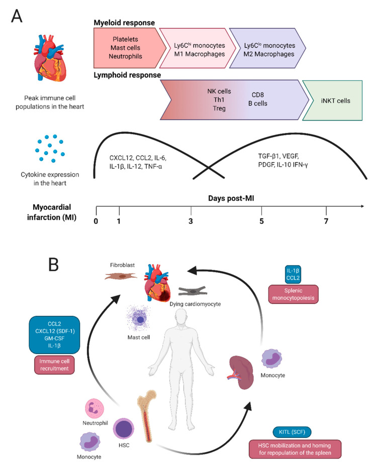
Figure 1.
Typical immune cell and tissue responses in the heart after permanent coronary artery occlusion. (A) The generalized pattern of immune cell infiltration to the heart after ischemic injury. Here, immune cell infiltrate is subdivided into myeloid and lymphoid populations. Myeloid cell infiltration spans approximately 1–3 days post-MI, while lymphoid cell populations infiltrate the heart at approximately 3–7 days post-MI. (B) Systemic tissue response to MI. After MI, signals from the heart (e.g., from degranulating mast cells, dying cardiomyocytes or activated fibroblasts) act on the spleen and bone marrow to initiate tissue repair. The activation of HSCs and migration of immune cells is triggered by a diverse array of molecules, including but not limited to CCL2, CXCL12 (SDF-1), GM-CSF and IL-1β. Monocytopoiesis is regulated by IL-1β and CCL-2. Gradually, HSCs in the bone marrow will repopulate the spleen in part through chemotactic molecules such as KITL (SCF).

