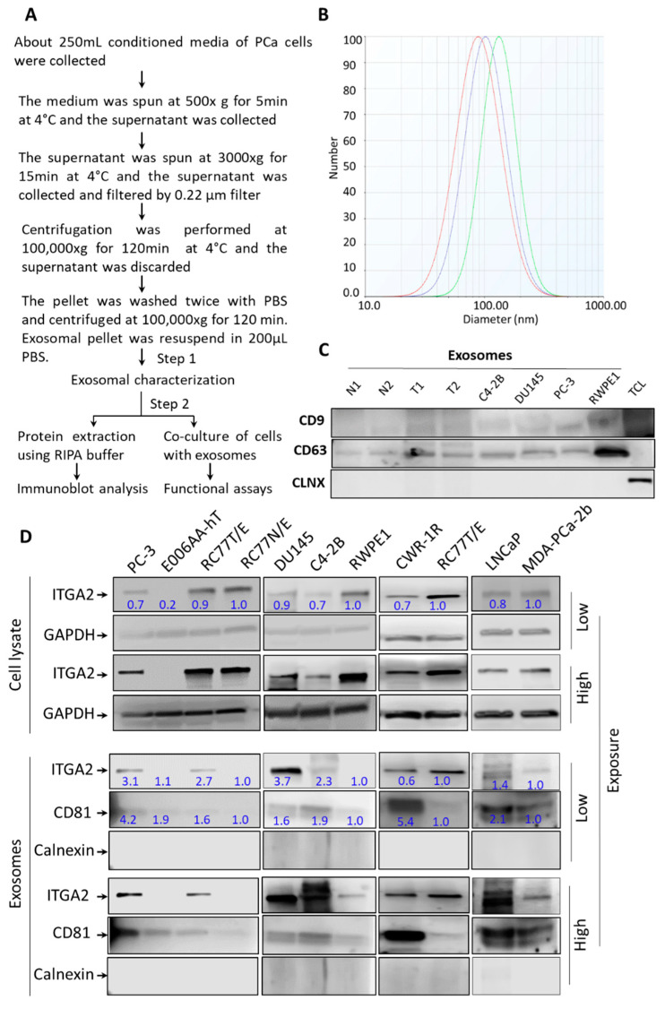
Figure 1.
Isolation, characterization and expression of ITGA2 in exosomes derived from PCa cells. (A). Schematic representation of exosome isolation from PCa cells by differential ultracentrifugation. Conditioned media collected from PCa cells were used for exosomes extraction. (B). Zeta potential analysis was performed to determine the number, average size, and homogeneity of exosomes isolated form of PC-3 cells (n = 3). (C). Exosomes were characterized by immunoblotting (IB) analysis. About 10 µg of exosomes derived from conditioned media of cells or plasma of PCa patients (T) and normal subjects (N) in addition to total cell lysate (TCL) of LNCaP cells were loaded onto 12.5% SDS-PAGE gel. IB analysis shows the expression of calnexin as a cellular marker and CD9 and CD63 as exosomal markers. (D). Enrichment of exosomal content of ITGA2 was evaluated in exosomes compared to cell lysates collected from LNCaP, C4-2B, DU145, PC-3, CWR-R1ca, RC77T/E, RC77N/E, RWPE-1, MDA-PCa-2b, and E006AA-hT cells using Western blot analysis. Twenty micrograms of protein lysates were loaded, and the membranes were incubated with anti-ITGA2, anti-GAPDH, anti-CD81, and anti-calnexin (CLNX) antibodies. These experiments were repeated at least three times.

