Figure 2.
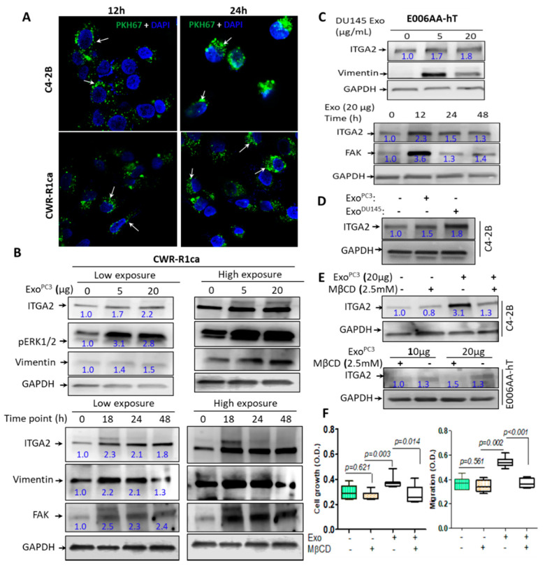
Exosomes-mediated transfer of ITGA2 in PCa cells. (A) Exosomal uptake of exosomes derived from PC-3 (ExoPC3, 10 µg/mL) labeled with PKH67 in C4-2B and CWR-R1ca cells. The green color of microbodies indicates exosomal uptake by recipient cells, and the blue color represents nuclear staining by DAPI. (B,C). Optimization of exosomes-mediated transfer in recipient cells. About 20 μg protein lysates were collected from CWR-1Rca (B) and E006AA-hT (C) cells, after their incubation with 0, 5, and 20 μg/mL ExoPC-3 for 48 h. Meanwhile, 20 μg protein lysate collected from CWR-1Rca and E006AA-hT cells previously incubated with 20 µg/mL ExoPC-3 for 18, 24, and 48 h. The membranes were probed with anti-ITGA2, anti-vimentin, anti-FAK and anti-pERK1/2 antibodies in addition to anti-GAPDH as a loading control protein. (D) Immunoblot (IB) analysis for C4-2B cells incubated with 20 μg/mL exoPC-3 for 24 h showing ITGA2 expression. (E) IB analysis for protein lysate collected from C4-2B and E006AA-hT cells treated without or with 2.5 mM MβCD in the presence of 20 µg/mL exoPC-3. (F) CWR-1Rca cells were treated with exoPC−3 with or without 2.5 mM MβCD for cell proliferation (72 h) and cell migration (24 h). The experiments were repeated at least twice. Data are statistically significant at p < 0.05.

