Figure 6.
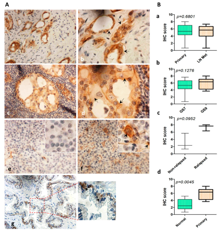
Expression of ITGA2 in PCa tumors with lymph node metastasis. Tissue cores were stained with anti-ITGA2 antibody. (A) Immunostaining of PCa tissue cores of primary (a, b) and lymph node metastasis tumors (c,d). Expression of ITGA2 was evaluated in non-relapsed (e), relapsed (f), and normal (g) tissue specimens. (B) Immunohistochemical score of ITGA2 in primary versus tumors with lymph node metastasis (a). High Gleason score (GS9) versus low Gleason score (GS7) (b). Relapsed versus non-relapsed tumors (c), and normal versus primary tumors (d). Magnification was 400× (a, c, e–g) and 1000× (b,d inserts).

