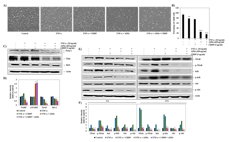
Figure 6.
The inhibitory effects of AIMs on MCF-7 cells treated with TNF-α and CDDP treatment. (A) the morphological representation of TNF-α treated cells with AIMs and CDDP. MCF-7 cells were pretreated with 400 µg/mL of AIMs 1 h at 37 °C, subsequently treated with or without 5 µg/mL of CDDP and 20 ng/mL of TNF-α at 37 °C, incubated for 48 h. (B) The cell viability is measured by the trypan blue exclusion assay. The data shown are mean ± SD of three different experiments performed separately. ** p < 0.05 non-treated versus treated group and *** p < 0.05 TNF-α treated versus AIMs and CDDP treated group. (C) Western blot analysis for cytotoxic effect of TNF-α, AIMs, and CDDP combined treatment for 48 h. The total lysates of MCF-7 cells with the above-mentioned treatment were resolved on SDS-polyacrylamide gels followed by transfer to PVDF membrane and probed with the specific primary and secondary antibody. The protein was visualized using chemidoc with the ECL detection kit. The data shown here are representative of at least three independent experiments. (D) The densitometry analysis of Western blot bands was normalized against actin and expressed as a mean of ± SD of at least three independent experiments # p < 0.05 TNF-α treated versus AIMs and CDDP treated group; (E) Western blot analysis of time dependent TNF-α treatment. MCF-7 cells were treated with 400 AIMs µg/mL of AIMs for1 h at 37 °C, subsequently treated with or without 5 µg/mL of CDDP and 20 ng/ml of TNF-α for 6 h and 12 h at 37 °C. The total lysates of MCF-7 cells with the above-mentioned treatment were resolved on SDS-polyacrylamide gels followed by transfer to PVDF membrane and probed with the specific primary and secondary antibody. The protein was visualized using chemidoc with the ECL detection kit. The data shown here are representative of at least two independent experiments. (F) The densitometry analysis of Western blot bands was normalized against actin and expressed as a mean of ± SD of at least three independent experiments # p < 0.05 TNF-α treated versus AIMs and the CDDP treated group. “+” and “−” represents the presence and absence of the compound, absence of the compound specified.

