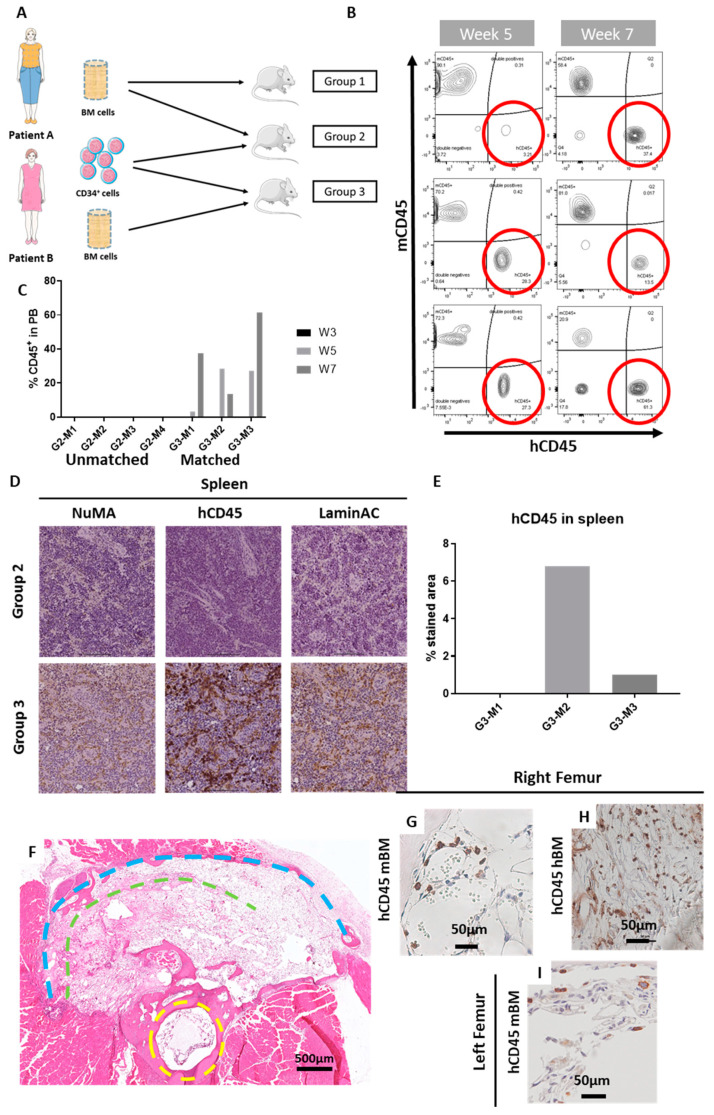
Figure 3.
CD34+ isolated from aged patients were able to establish a human haematopoietic system in the mice. Overview of the in vivo study. Group 1 (n = 4 mice) did not receive any CD34+ cells, whereas Groups 2 (G2) and 3 (G3) were implanted with 85,000 CD34+ cells isolated from Patient B. Group 3 (n = 3 mice; G3-M1, G3-M2, G3-M3) had matched cells from the same patient, and Group 2 (n = 4 mice; G2-M1, G2-M2, G2-M3, G1-M4) had cells from different patients (A). Flow cytometry was utilised to monitor cell engraftment. After 5 weeks (B), hCD45+ cells were detectable in peripheral blood of the mice, with values ranging from 3.21% to 27.3%. Flow cytometry at week 7 verified their engraftment and was increased in some cases (C). Immunohistochemical analysis revealed the presence of hCD45+ cells in the spleen of the mice that receives the BM and CD34+ cells from the same patient (D), with up to 6% of hCD45+ stained area (E). H&E staining of a cross section of a mouse leg, with the yellow dashed line indicating the mouse femur and the green and blue representing the inner and outer implanted scaffolds, respectively (F). Further immunohistochemical analysis assisted to locate hCD45+ cells in the right femur of the mice (G–H), in both the murine BM compartment (G) and in the hBM compartment (H). hCD45+ cells also migrated to the contralateral, non-operated leg (I).

