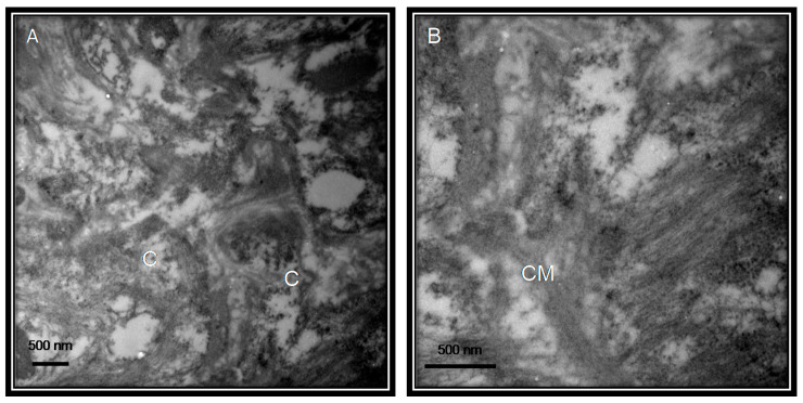
Figure 2.
Postmortem lung biopsy of a 28 year old male patient with acute myocardial infarction and aortic stenosis as control subject. The bronchiole without viral particles. The pictures of the electron microscopy panels (A,B) were made at 25,000× and 50,000× respectively with a Jeol JEM-1011 electronic microscope. Abbreviations: C = cellule, CM = cellular membrane.

