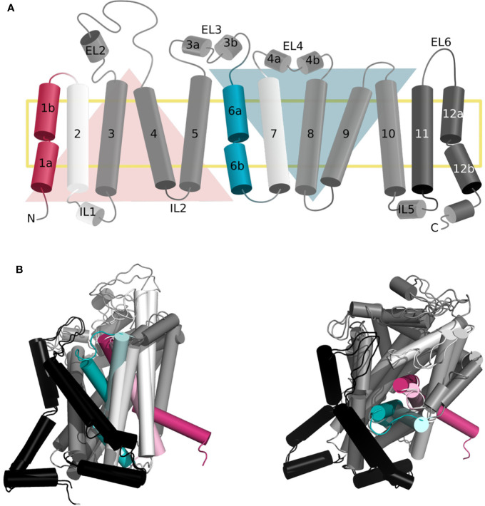
Figure 1.
Topology of the gated pore mechanism of transport. (A) Topology of hSERT. The transmembrane helices constituting the bundle and scaffold domain are shown in white and dark gray, respectively, with the exception of the gating helices TM1 and TM6 in red and cyan. The two inverted repeats are indicated by two triangles, while the two additional helices TM11 and TM12 are shown in black. This figure was adapted from ref (Hellsberg et al., 2019). (B, C) Superposition of the three-dimensional structures of hSERT in outward open [PDB ID 6DZY (Coleman et al., 2019)] and inward open [PDB ID 6DZZ (Coleman et al., 2019)] conformations from the side (B) and top (A) view, with the same color code as panel (A) the gating helices in the outward open conformation are shown in light red and light cyan, to contrast with the inward open state.

