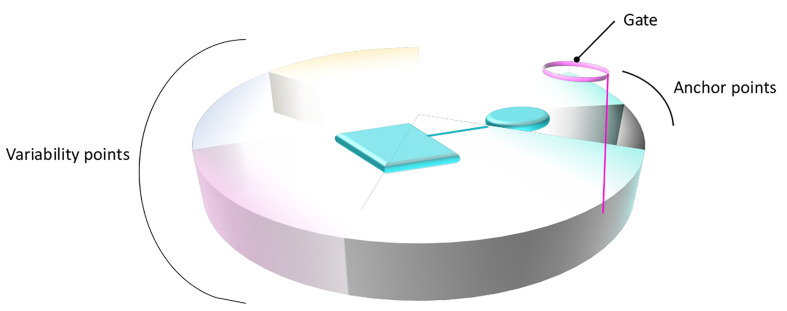
Figure 4.
General structural annotation of the primary binding site of LeuT fold transporters. Gathering the information available from the distinct studies on the SLC6 and SLC7 members permits to structurally and functionally map the binding site of LeuT fold transporters. The binding site is symbolized as a disc divided into distinct portions that constitute the binding site, following the same color code as in Figure 1. The functional annotation of each area is indicated on the side of the disc. A bound ligand is represented in cyan. The anchored part of the ligand, conserved within subgroups is shown as a cylinder. The part of the ligand allowing variability and conferring various transport activities is shown as a square.

