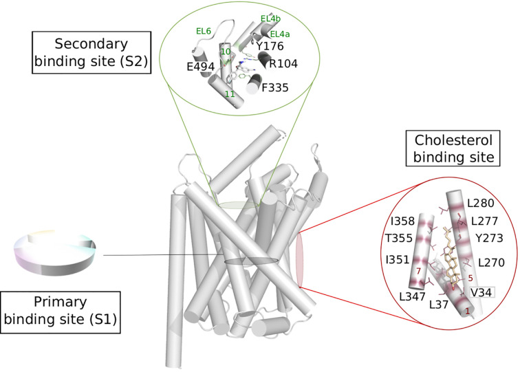
Figure 5.
Possible functional mapping of the LeuT fold. Building on the functional mapping of the binding site proposed in Figure 4, we show here two additional area where functional mapping could be beneficial, i.e., on the secondary and cholesterol binding sites. The residues involved in binding are shown in sticks and labeled (hSERT numbering for the secondary site and dDAT numbering for the cholesterol binding site), the helices and loops are named on green and red on the secondary et cholesterol binding site, respectively. Expanded to the whole fold, a systematic annotation could help the identification of new sites that could be targeted in ligand discovery.

