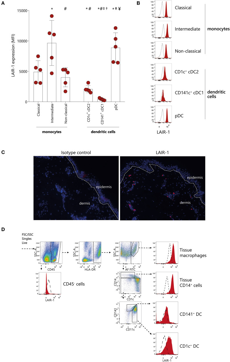
Figure 1.
LAIR-1 is differentially expressed on circulating monocytes subsets and dendritic cells subpopulations and on skin immune cells. (A) Quantification and (B) representative histograms of LAIR-1 expression, represented as median fluorescence intensity (MFI), on classical, intermediate and non-classical monocytes as well as on CD1c+cDC1s, CD141+cDC2s, and pDCs, determined on peripheral blood mononuclear cells (PBMC) by flow cytometry. Results are represented as mean with SD. Differences were considered statistically significant when p < 0.05: *vs. classical monocytes, #vs. intermediate monocytes, †vs. non-classical monocytes, ‡vs. CD1c+cDC1s, ¥vs. CD141+cDC2s (one-way ANOVA test). (C) Immunofluorescence analysis of LAIR-1 (red staining), in normal skin section and isotype control is shown as negative control. DAPI nuclear counterstain is shown in blue. Representative images out of three independent stainings were acquired in 20 × magnification. (D) Flow cytometry of enzymatically digested skin. Gating strategy used to identify tissue macrophages, tissue CD14+ cells, CD141+, and CD1c+ DCs is shown. LAIR-1 expression (filled) on these cells is shown compared to isotype control (dashed). Representative data from three donors are shown.

