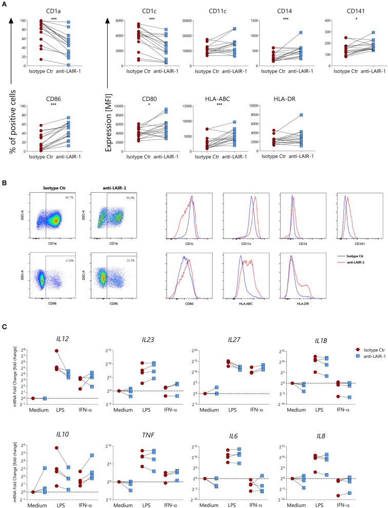
Figure 5.
LAIR-1 activation during differentiation of monocyte-derived dendritic cells results in phenotypic and cytokine profile alterations. (A) Purified monocytes were differentiated for 6 days into monocyte-derived dendritic cells using GM-CSF and IL-4 in the presence of anti-LAIR-1 agonist (Dx26) or isotype control and the expression of CD1a, CD1c, CD11c, CD14, CD141, CD86, CD80, HLA-ABC, and HLA-DR was assessed by flow cytometry. Quantification is shown as percentage (%) of positive cells or median fluorescence intensity (MFI). (B) Representative plots or histograms are shown. (C) Monocyte derived dendritic cells differentiated in the presence of anti-LAIR-1 agonist (Dx26) or isotype control were stimulated during 4 h with TLR4 agonist- LPS or IFN-α and the IL12A, IL23A, IL27A, IL1B, IL10, TNF, IL6, IL8 gene expression was evaluated by qRT-PCR. Results are represented as paired samples. Statistically significant differences were considered when *p < 0.05, ***p < 0.001 (Wilcoxon's test).

