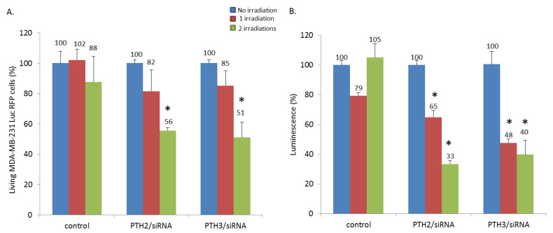
Figure 4.
(A) MDA-MB-231 Luc RFP cells were incubated with PTH2/siRNA and PTH3/siRNA at a P+/P- ratio of 100 during 24 h. The cells were irradiated for 5 min using a standard fluorescent microscope with a mercury lamp at 450 nm, magnification x4. After 24 h, the cells were irradiated a second time with the same process. The day after, the cells were submitted to an MTT assay to quantify the cell death. (B) Luciferase activity assay showing the transfection of a 21-mer siRNA targeting the expression of luciferase inside MDA-MB-231 Luc RFP cells. The experiments were carried out with PTH2/siRNA and PTH3/siRNA at a P+/P- ratio of 100 without or after one or two irradiations (450 nm, 5 min). * Statistical significance (p < 0.05), of the irradiated condition versus non irradiated condition using Student’s t-test.

