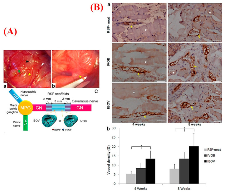
Figure 5.
(A) Macroscopic views representing the implantation of regenerated silk fibroin (RSF) scaffolds into a rat animal model: (a) exposed major pelvic ganglion (MPG), pelvic nerve (PN), hypogastric nerve (HN), and cavernous nerve (CN) are marked as black, green, white, and yellow arrows, respectively; (b) creating the CN gap (arrow) (length of 5 mm) by scissors; and (c) schematic illustration of suturing process of RSF scaffolds and two CN ends. (B) Immunohistochemistry (IHC) staining of the harvested samples for evaluating new vessels in the retrieved scaffolds of RSF-neat, inner-VEGF/outer-brain-derived neurotrophic factor (BDNF) (IVOB), and inner-BDNF/outer-VEGF (IBOV) after 4 and 8 weeks of implantation (a), in which the asterisk (*) shows the scaffold fragments and the yellow arrows show vessels (bar: 100 μm); and (b) graph showing the vessel densities in (a) in which the asterisk (*) shows significant differences among the groups at each time point (p < 0.05). Adapted from Zhang et al. [274], with permission from American Chemical Society.

