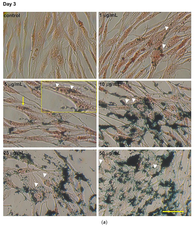
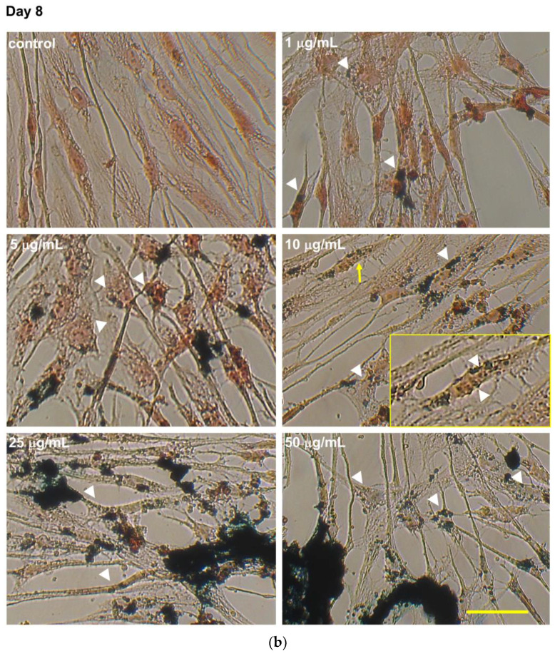
Figure 4.
Intracellular evaluation of Fe3O4NPs by Perls’ Prussian blue staining in hNLCs cultured with Fe3O4NPs. Representative micrographs, by light microscopy, of hNLCs after 3 days (a) and 8 days (b) in neurogenic medium with increasing concentrations of Fe3O4NPs (1–50 μg/mL). Prussian blue staining showed fine intracellular blue spots of Fe3O4NPs around the nucleus already at the lowest concentration tested (1 μg/mL). The intracellular iron accumulation increased with increasing concentration and the aggregations/agglomerations of Fe3O4NPs were also visible extracellularly. White heads indicate intracellular nanoparticles and yellow arrows indicate the magnifications (2X) of the areas of the insert selection. Scale bar: 100 μm.


