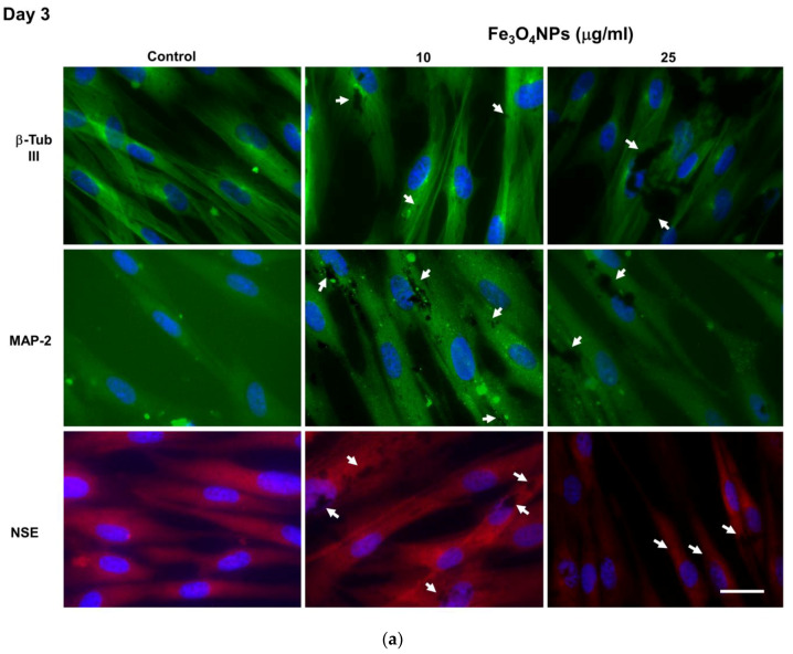
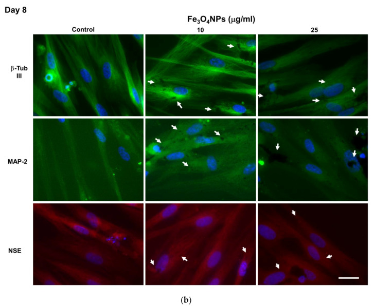
Figure 7.
Neuronal marker protein expressions in hNLCs cultured with Fe3O4NPs. Fe3O4NPs apparently affected the transdifferentiation of CL-hMSCs starting from 25 μg/mL: hNLCs at day 3 (a) and day 8 (b) of transdifferentiation exhibited a decrease of fluorescence intensity of neuron markers such as β-Tub III (green fluorescence), MAP-2 (green fluorescence) and NSE (red fluorescence). Black spots of Fe3O4NPs (intracellularly and on the cell membrane) were also visible. Nuclei were stained with Hoechst 33258. White arrows indicate Fe3O4NPs (dark spots). Scale bar: 100 μm.


