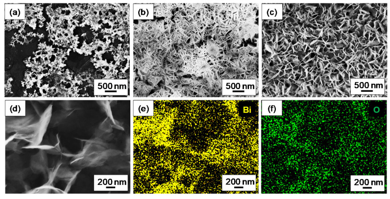
Figure 2.
Scanning Electron Microscopy images of (a) GP_Bi at centre, (b) GP_Bi at edge of active area and (c) GP_naf_ex_Bi at edge of active area; (d) Scanning Electron Microscopy image and (e,f) corresponding chemical maps obtained by Energy Dispersive X-ray Analysis of bismuth and oxygen of GP_Bi at the edge of the active area.

