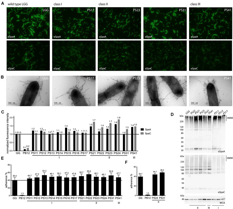
FIGURE 1.
Phenotypic assessment of piliation and mucus adhesion of highly mucus-adherent derivatives of L. rhamnosus GG. (A) Immunofluorescent labeling of SpaA and SpaC on cell surface of derivatives. Strains were labeled with either SpaA or SpaC antisera and detected with Alexa 488-labeled secondary antibody. Representative images are included for each strain. (B) Immunoelectron micrographs of strains double labeled with SpaC and SpaA antibodies. Both SpaC (5 nm gold particles) and SpaA (10 nm gold particles) are imaged on the pili of the different derivative strains (a representative figure is shown for each strain). (C) Quantitative immunofluorescent labeling results of SpaA and SpaC. Fluorescent signals of strains labeled with either SpaA and SpaC (detected by Alexa 488-labeled antibodies) were normalized to DAPI fluorescence. These values were further normalized to the fluorescence of the parental L. rhamnosus GG strain (indicated with a line to facilitate interpretation). A representative experiment is depicted. (D) Western blot analysis of cell wall associated proteome of each derivative. The pilus ladder (HMW for High Molecular Weight fraction) of the derivatives was visualized by probing cell wall extracts of each strain with SpaA and SpaC antibodies. Total amount of proteins is shown as a reference with a whole cell antibody (WCA) blot. (E) Mucus adhesion of the derivatives. The average result of 3 biological measurements (with each 3–6 technical repeats) is depicted for each derivative with wild-type L. rhamnosus GG as a positive control and the non-piliated derivative PB12 as a negative control. Standard error of mean (SEM) is shown. (F) Adhesion to human mucus. Adhesion of two derivatives, together with the positive and negative controls, to mucus isolated out of a human specimen, to validate porcine results. The average of three biological repeats is depicted with standard error of mean (SEM).

