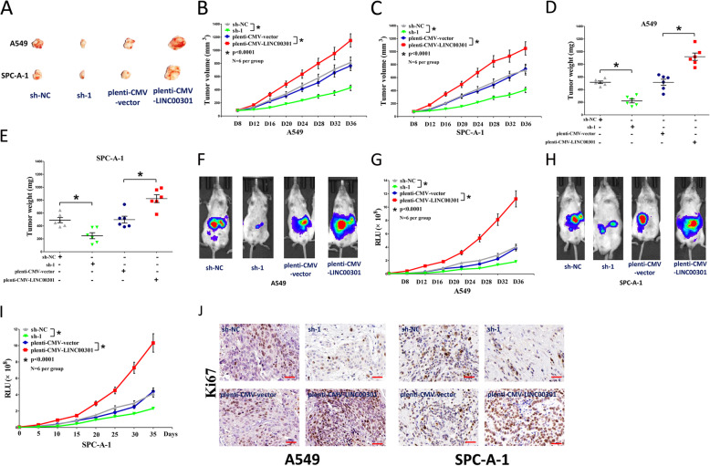
Fig. 3.
LINC00301 promotes tumor growth in vivo. a Representative images tumors separated from nude mice. b, c Tumor volume was measured in nude mice. d, e Tumor weight was examined in nude mice. Each group contained six mice (n = 6). f–i Representative bioluminescence imaging (BLI) images and quantification of BLI in the tumor regions for nude mice. Data were shown as the mean ± SD; * p < 0.05, in comparison to the sh-NC or pLenti-CMV-control group. j Representative images of Ki-67 staining in tumors isolated from the nude mice. Bar = 50 μm. Assays were conducted in triplicate. *p < 0.05, means ± SD was shown. Statistical analysis was subjected to Student’s t-test

