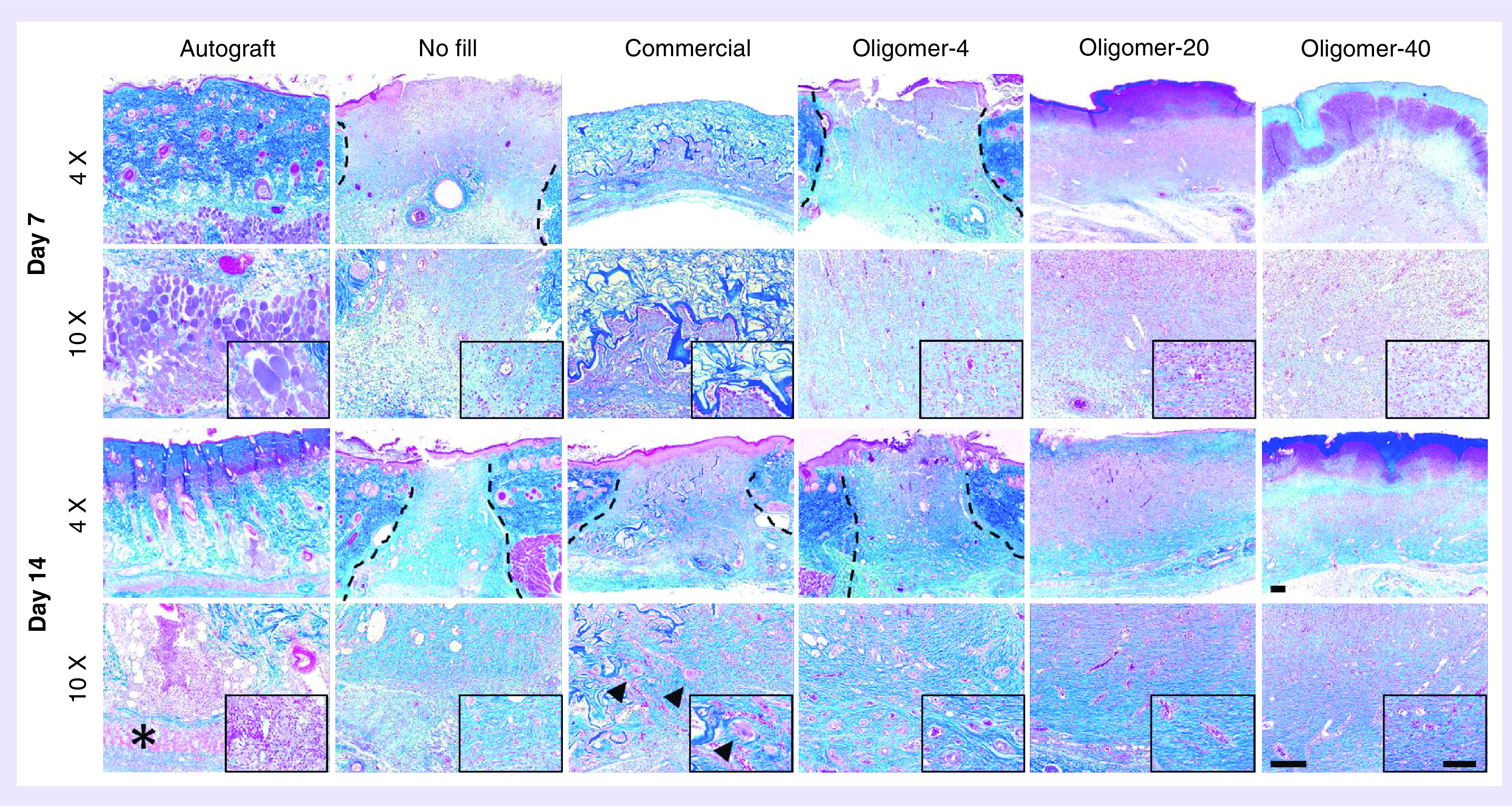
Figure 5. . Histological cross-sections (4× and 10× with 40× inset) of excised skin wounds 7 days (above) and 14 days (below) stained with Masson’s trichrome following treatment with autograft, no fill, commercial dressing, Oligomer-4, Oligomer-20 and Oligomer-40.

Images represent center region of wound with inset focused on cellular response. Asterisks denote atrophying panniculus carnosus muscle, arrowheads denote giant cells and dashed lines indicate wound borders if visible. Scale bars: 4×: 200 μm; 10×: 200 μm; 40×: 100 μm.
