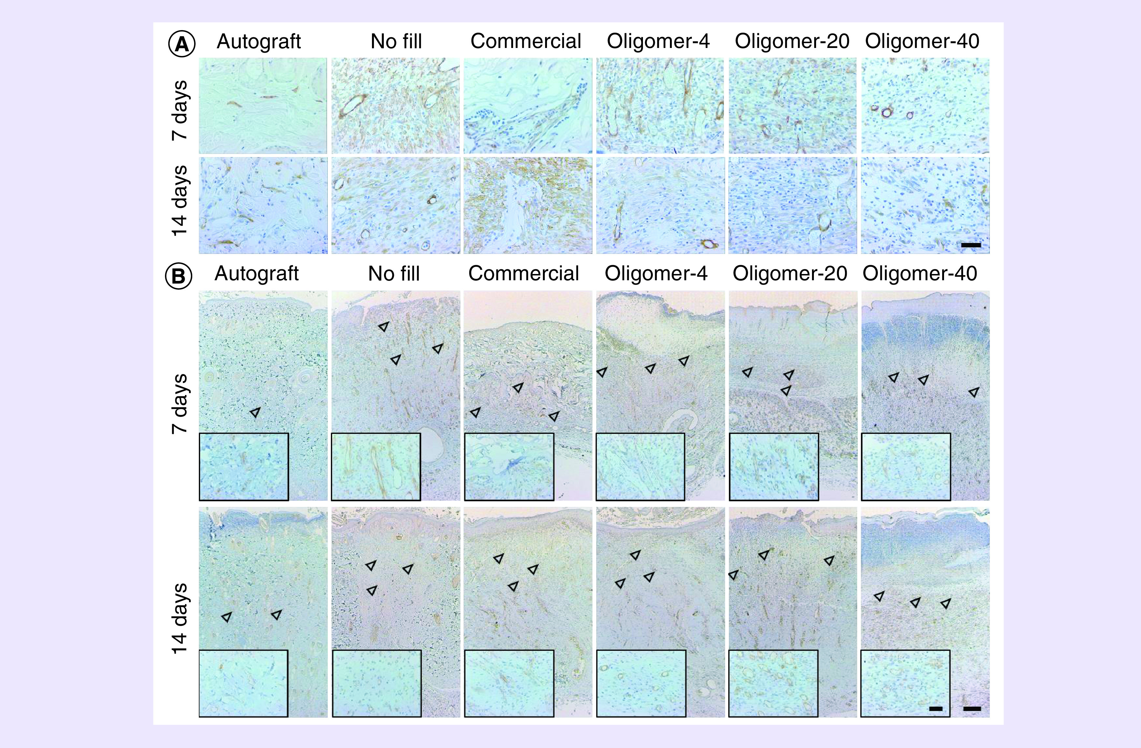
Figure 6. . Time-dependent changes in cellularization and vascularization of wounds following treatment with autograft, no fill, commercial dressing, Oligomer-4, Oligomer-20 and Oligomer-40.

(A) Histological cross-sections of excised wound tissues stained for blood vessel and myofibroblast marker α-SMA (brown; 7 and 14 days) and (B) endothelial cell marker CD31 (brown; 7 and 14 days). Arrowheads denote presumed level of vascularization based on presence of CD31 positive stained lumens with identifiable red blood cells. Scale bars: (A) 50 μm; (B) 200 μm, (B), inset: 50 μm.
