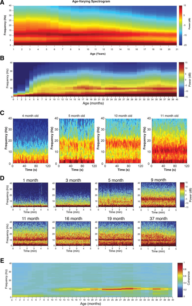
Figure 4.

Combined spectrograms of propofol-induced frontal EEG activity for patients 1–21 y of age. Prominent alpha and slow oscillations are present across the entire age range in spite of shifts in overall power that are dependent on age (A). Combined spectrograms of sevoflurane-induced, frontal EEG activity for patients 0–40 mo of age (B). Four individual spectrograms of frontal EEG power from infants receiving propofol anesthesia across the first year of life (C). Individual spectrograms of frontal EEG power from infants receiving sevoflurane anesthesia across the first 3 y of life (D). High-frequency power is largely absent in 0- to 4-mo-old infants, but an alpha/beta oscillation emerges around 9 mo that travels downward in peak frequency until around 1 y of age (C, D). Combined coherograms of sevoflurane-induced, frontal EEG activity for patients 0–40 mo of age (E). Only recordings when patients were maintained at surgical levels of anesthesia are presented. EEG indicates electroencephalogram. Figure panels B, D, and E were adapted from the British Journal of Anaesthesia, 120, Cornelissen L, Kim SE, Lee JM, Brown EN, Purdon PL, Berde CB, “Electroencephalographic Markers of Brain Development During Sevoflurane Anaesthesia in Children up to 3 years old,” 1274–1286, 2018, with permission from Elsevier.33 Figure panels A and C were adapted from Lee JM, Akeju O, Terzakis K, et al, “A Prospective Study of Age-Dependent Changes in Propofol-Induced Electroencephalogram Oscillations in Children,” Anesthesiology, 2017, 127, 2, 293–306.32
