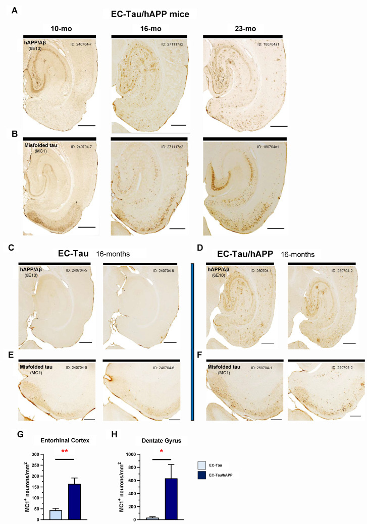
Fig 1. hAPP/Aβ-associated acceleration of tau pathology along the EC-HIPP network.
The EC-Tau/hAPP mouse line was created to model functional interactions between hAPP/Aβ and hTau pathologies in a well-characterized neuronal circuit in vivo (EC-HIPP network). A-B. Horizontal brain sections from a sample of 10-, 16-, and 23-month-old EC-Tau/hAPP mice were processed for immunohistochemical detection of hAPP/Aβ (6E10, anti-beta amyloid) and abnormal, misfolded tau (MC1, conformationally dependent). A clear, age-dependent progression of Aβ and tau pathology within the EC-HIPP network was evident. Importantly, MC1+ immunostaining revealed increased pathological tau within hippocampal neurons at 16 months compared with 10 months of age. Scale bars, 500 μm. C-D. To determine whether hAPP/Aβ aggravates tau accumulation and accelerates pathological tau spread at the 16-month time point, horizontal brain sections from EC-Tau/hAPP mice (n = 6 total; n = 3 female, n = 3 male) and age-matched EC-Tau littermates (n = 5 total: n = 3 female, n = 2 male) were processed for 6E10+ and MC1+ immunostaining. Representative, adjacent brain sections from 2 mice sampled are shown for both 6E10 and MC1. The 16-month EC-Tau/hAPP mice exhibited robust Aβ accumulation and plaque deposition throughout the EC and HIPP. Diffuse Aβ accumulation comprised the majority of the pathology in these regions, with occasional small, compact plaques and large, dense-core Aβ plaques present. EC-Tau mice did not exhibit 6E10+ immunoreactivity. Scale bars, 500 μm. E-F. MC1+ immunostaining revealed an acceleration of tau pathology in the EC of EC-Tau/hAPP mice, characterized by an increased number of neurons with abnormally conformed tau localized within somatodendritic compartments. Scale bars, 250 μm. G-H. Semiquantitative analysis of MC1+ cell counts was performed in the EC and DG of both EC-Tau/hAPP and EC-Tau mice. Mean MC1+ cell counts (MC1+ neurons/mm2) in EC-Tau/hAPP brain sections were over 4-fold and over 20-fold greater than EC-Tau brain sections in the EC and DG, respectively. EC: EC-Tau/hAPP, 164.02 ± 27.12 versus EC-Tau, 43.23 ± 9.35. DG: EC-Tau/hAPP, 631.59 ± 212.90 versus EC-Tau, 30.95 ± 14.20. Unpaired t-tests with Welch’s correction: EC, t = 4.211, p < 0.01; DG, t = 2.815, p < 0.05. Graphs and numerical values in figure legend represent mean ± SEM for the averaged MC1+ neurons/mm2 values from 3 independently processed brain sections per mouse. *p < 0.05; **p < 0.01. Source data are available in S1 Data. Aβ, amyloid-beta; DG, dentate gyrus; EC, entorhinal cortex; hAPP, human amyloid precursor protein; HIPP, hippocampus; hTau, human tau; MC1, antibody for misfolded tau; SEM, standard error mean.

