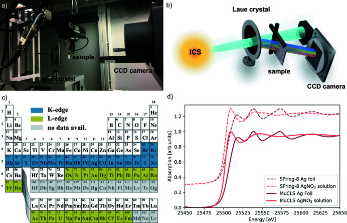
Figure 11.
The XAS set-up is depicted in (a) with a schematic of the beam path shown in (b). The elements whose K- and L-edges can be covered by the energy range of the ICS of the MuCLS are highlighted in (c) in blue and green, respectively. Elements for which no data are available from the NIST database (NIST, 2005 ▸) are coloured in grey. As an example, a measurement of a silver foil and a silver nitrate solution is depicted in (d). For comparison, synchrotron measurements of the same substances are included and shifted upwards.

