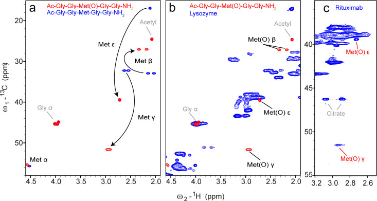
Figure 2.
1H–13C HSQC spectra of the reference peptides for the detection of Met(O), lysozyme, and H2O2-treated rituximab. (a) Overlay of the reference spectra of peptides Ac–Gly–Gly–Met(O)–Gly–Gly–NH2 (red) and Ac–Gly–Gly–Met–Gly–Gly–NH2 (blue) under denaturing conditions (7 M urea, pH 2.3). (b) Overlay of the spectra of the reference peptide Ac–Gly–Gly–Met(O)–Gly–Gly–NH2 (red) and denatured lysozyme (blue), to identify unique chemical shifts of Met(O) that differ from the random-coil chemical shift correlations of the 20 natural amino acids. (c) 1H–13C HSQC spectrum of treated (0.35% H2O2 for 30 min at RT) rituximab (512 × 512 complex points, 104 scans, a recycle delay of 3 s, total measurement time of 2 days and 14 h) under denaturing conditions (7 M urea-d4 in D2O) at pH 2.3.

