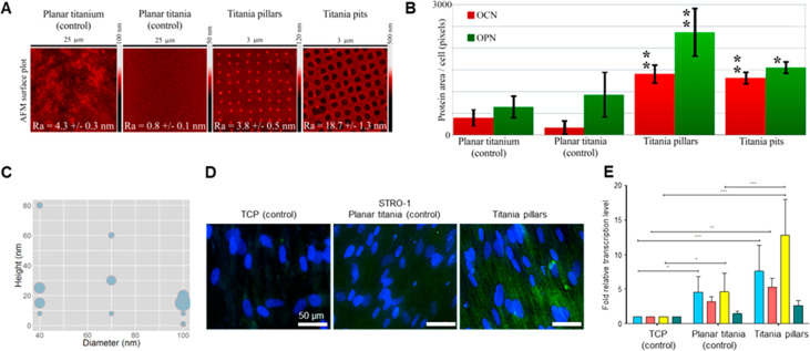
Figure 2.
(A) AFM images of the tested surfaces with superimposed image Ra ± standard deviation. The samples represent polished titanium, planar TiO2 surface derived from sintered sol–gel, NSQ nanopillars produced in sol–gel-derived TiO2 with 100 nm diameter, 15–25 nm tall pillars, and NSQ nanopits produced in sol–gel-derived TiO2 with 200 nm diameter, 60 nm deep pits. (B) OCN and OPN fluorescence levels from STRO-1 SSCs 21-day in vitro culture on the surfaces of panel A. (C) Bubble plot depicting the production of OCN for different pillar diameters and heights. (D) OPN immunofluorescence using a selective antihuman OPN antibody in STRO-1 SSCs after 21 days of in vitro culture. Note enhanced OPN protein expression on nanopillars in comparison to TCP and Titania planar substrates (blue–cell nuclei, green – OPN protein). (E) Real-time qPCR analysis of ALP (blue), collagen 1 (red), OPN (yellow), and OCN (green) in STRO-1 SSCs cultured in vitro on test surfaces and tissue culture plastic for 21 days. STRO-1 TCP is taken as a negative control. Results expressed as mean ± SD, triplicate samples, individual experiment repeated five times, 2-way ANOVA test, *p < 0.05, **p < 0.01, ***p < 0.001.

