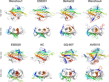
Fig. 3. Three-dimensional structure of coat protein dimers.

Proteins representing different variations of the CP fold are shown with one of the monomers rainbow-colored blue (N terminus) to red (C terminus) and the other monomer shown in light gray. All CP dimers are shown as seen from outside of the particle (top view) and in a roughly perpendicular view looking lengthwise down the C-terminal α helices (side view).
