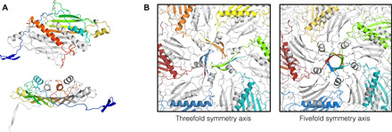
Fig. 4. The unusual coat protein of bacteriophage Beihai14.

(A) Three-dimensional structure of the Beihai14 CP. The CP dimer is colored and shown in two different orientations as in Fig. 3. (B) Interactions around the threefold and fivefold icosahedral symmetry axes in the assembled particle. The CP monomers with their N-terminal extensions involved in the interactions are shown in color.
