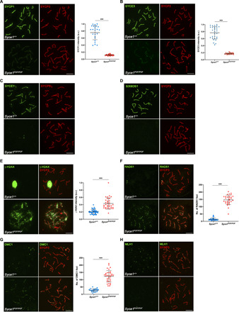
Fig. 2. Syce1POF/POF spermatocytes are not able to synapse and DSBs are deficiently repaired.

(A) Double immunolabeling of WT pachytene and Syce1POF/POF zygotene-like spermatocytes with SYCP3 (red) and SYCP1 (green). In Syce1POF/POF spermatocytes, AEs fail to synapse and show a weak staining of SYCP1 along the axial elements (AEs). a.u., arbitrary units. (B to D) Double immunolabeling of spermatocyte spreads with SYCP3 (red) and the CE proteins (green). Syce1POF/POF zygotene-like spermatocytes showed a highly reduced signal of SYCE3 (B) and the absence of (C) SYCE1 and (D) SIX6OS1 from the AEs. (E) Double immunolabeling of γ-H2AX (green) and SYCP3 (red) in spermatocyte spreads from WT and Syce1POF/POF mice. γ-H2AX staining was persistent in Syce1POF/POF zygotene-like spermatocytes, but was restricted to the sex body in WT pachytene cells. (F and G) Double immunofluorescence of (F) RAD51 or (G) DMC1 (green) and SYCP3 (red). Syce1POF/POF zygotene-like spermatocytes showed increased numbers of foci of RAD51 and DMC1 along the AEs in comparison with WT, indicating unrepaired DSBs. (H) Double immunolabeling of MLH1 (green) and SYCP3 (red) showing the absence of COs (MLH1) in arrested Syce1POF/POF spermatocytes. Fluorescence intensity levels (A, B, and E) and number of foci (F and G) from WT and zygotene-like arrested spermatocytes are quantified in the right-hand plots. Welch’s t test analysis: ***P < 0.0001. Scale bars, 10 μm.
