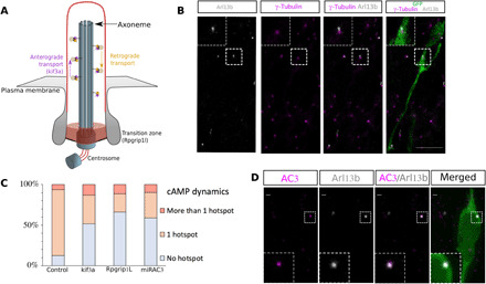
Fig. 3. PC ablation and AC3 KD lead to disappearance of the cAMP hotspot in the majority of neurons.

(A) Scheme of the PC. (B) Immunohistochemistry of a GFP-positive RMS neuron (green) in control Kif3alox/lox mice showing a short Arl13b-positive PC (gray) connected to the γ-tubulin–positive centrosome (magenta). The PC length was, on average, 0.64 ± 0.03 μm (N = 3 mice, n = 35 PC). Scale bar: 10 μm. (C) Percentage of neurons with no hotspot, one or more than one hotspot in control or Kif3alox/lox and Rpgrip1lox/lox CRE, or mirAC3-transfected neurons. Control 82% cells with hotspot (N = 10, n = 101) versus CRE-Kif3alox/lox 30% (N = 4, n = 36) and CRE-Rpgrip1llox/lox 20% (N = 7, n = 52) and miRAC3 31% (N = 3, n = 62). (Fisher’s exact test, P < 0.001). (D) Immunocytochemistry of a GFP-positive neuron (green) in control Kif3alox/lox mice showing AC3 subcellular localization (magenta) in the Arl13B-positive PC (gray). Scale bars: 1 μm
