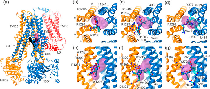
Figure 8.
Docking of pentapeptides to the SUR1 cavity. (a) General view of the SUR1 structure with the KN tail peptide shown in black and the approximate binding position of GBM63 shown as a pink volume. (b–d) KNt26, KNt37, and KNt48 pentapetide binding position in a top view, and (e–g) side view, with the main residues forming the binding spaces shown in a cyan (TMD1) and orange (TMD2) stick representation.

