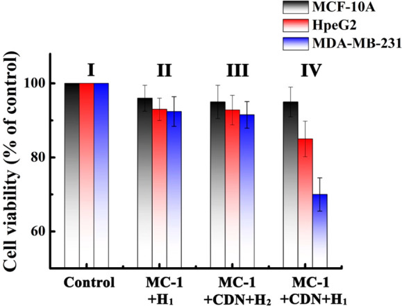
Figure 4.

Cytotoxicity of the CDN “S”/H1/MC-1 toward MCF-10A, HpeG2, and MDA-MB-231 cells and appropriate control systems. Panel I: no addition of the components to the cells. Panel II: MC-1 and H1 were incorporated in all kinds of kinds of cells (No CDN added). Panel III: MC-1, the CDN “S”, and H2 were incorporated into the cell (no H1). Panel IV: MC-1, CDN “S”, and H1 were incorporated into all three kinds of cells. The cell viability was monitored after a time interval of 2 days at the respective conditions. Error bars derived from N = 3 experiments.
