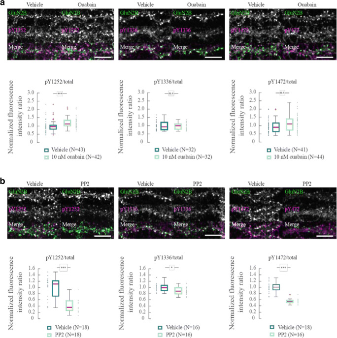
Fig. 4.
Tyrosine phosphorylation of GluN2B. a Antibody labeling of the three SFK phosphorylation sites of GluN2B: pY1252, pY1336, and pY1472, and total GluN2B in dendrites following 5 min application of vehicle or 10 nM ouabain. Boxplots of the ratio of the fluorescent intensity of pYGluN2B/GluN2Btotal normalized to the mean under control conditions with vehicle. b As in a but exposed to DMSO (vehicle) or 10 μM PP2 for 5 min. Boxplots of the ratio of the fluorescent intensity of pYGluN2B/GluN2B total normalized to the mean under control conditions with DMSO. n.s.—p > 0.05, *p < 0.05, ***p < 0.001, Wilcoxon rank sum test. Scale bar = 10 μm

