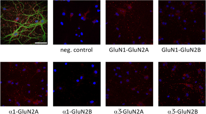
Fig. 5.
Proximity ligation assay (PLA) images of rat hippocampal neurons. PLA was performed using antibodies against Na,K-ATPase α-subunits (NKAα1 and NKAα3) and against GluN2-subunits (GluN2A and GluN2B). Omission of primary antibodies was used as negative control, and antibodies against GluN1 was used as a positive control. Red dots indicate PLA signal; cell nuclei are identified using DAPI stain. Upper left image is PLA of NKAα3 with GluN2B counterstained with pan neuronal marker in green to visualize neurons with extensions. Scale bar = 50 μm

