
Fig. 7.
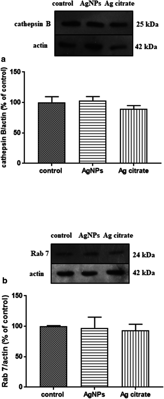
Expression of proteins involved in lysosomal functions during autophagy in brain homogenates obtained from rats exposed to control (saline), silver nanoparticles (AgNPs), and silver citrate; representative immunoblots for protein levels of cathepsin B (a) and Rab 7 (b). The graphs illustrate the results of densitometric measurements of 4 independent immunoblots performed using 4 distinct animals, expressed as a percentage of control. The relative protein density was measured against β-actin as an internal standard. The values represent the means ± SD; P > 0.05 was considered not significantly different vs. control group (one-way ANOVA with post hoc Dunnett’s test)