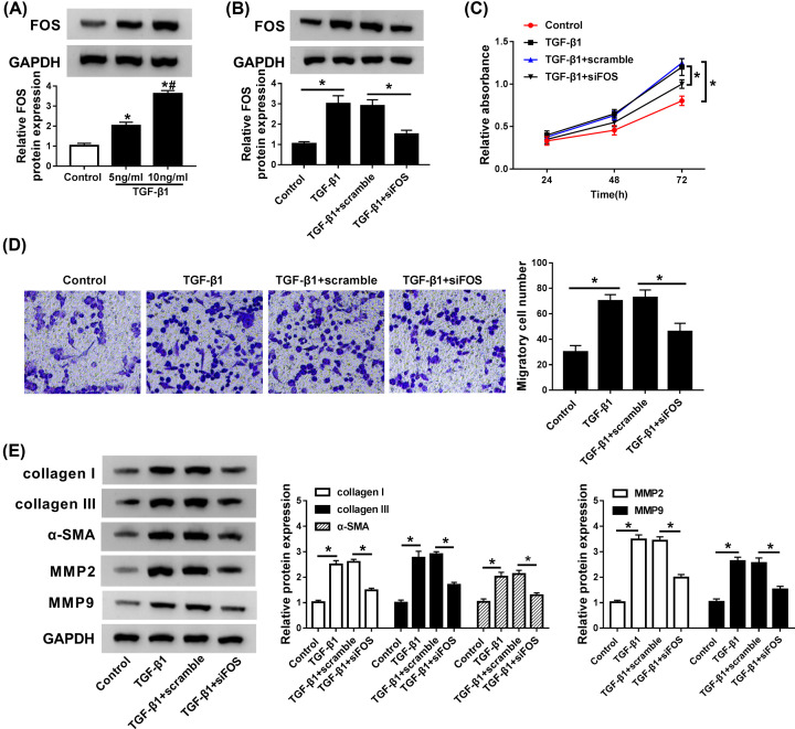
Figure 5. FOS knockdown overturned the stimulatory effects of TGF-β1 on cell proliferation, migration, and myofibroblast differentiation in CFs.
(A) CFs were stimulated with different concentrations (0, 5, 10 ng/ml) of TGF-β1 for 24 h, and FOS protein expression was detected by Western blot assay. (B) CFs treated with TGF-β1 (10 ng/ml) were transfected with scramble or si-FOS, followed by the detection of FOS protein expression by qRT-PCR. (C and D) Cell proliferation and migration ability were assessed by CCK-8 and Trans-well assays. (E) The abundances of collagen I, collagen III, α-SMA, MMP2, and MMP9 proteins were measured by Western blot assay. *P<0.05 compared with corresponding control, #P<0.05 compared with TGF-β1 (5 ng/ml).

