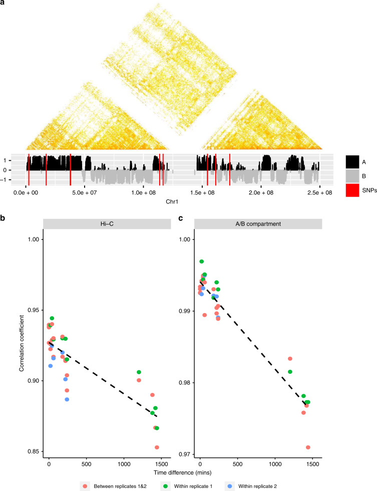
Fig. 2. Illustration of Hi-C dynamics.
a Hi-C interaction matrix (100 kb resolution) of replicate 1 for chr1 at time 0 min (upper) with corresponding A/B compartments (lower), where red lines represent positions of SNPs. b Correlation changes between Hi-C data with respect to differences between times, where the dashed line is the fitted linear line for the correlation coefficients. c Correlation changes of A/B compartments with respect to the differences between times, where the dashed line shares the same information as conveyed in the plot b.

