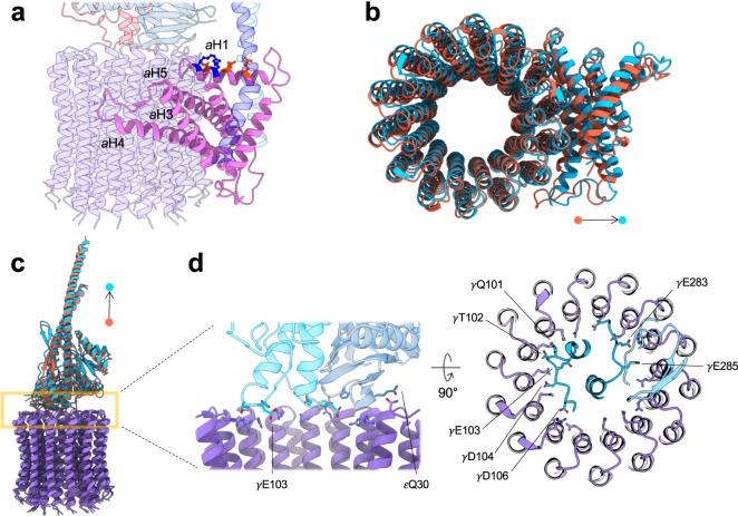
Fig. 4. Interaction of the γ-ε central shaft with the membrane FO domain.
a Spatial arrangement of the subunit a in the membrane. Color codes: subunit a (light pink), c14 ring (purple), bb′ stator (blue and light blue), γ subunit (crimson), and ε subunit (steel blue). Charged residues on the aH1 are shown as a ball-and-stick model. Negatively (aGlu73, aGlu77, and aAsp81) and positively (aArg80 and aLys84) charged residues are shown in orange and blue, respectively. b Alignment of the membrane c-ring rotors of the reduced and oxidized states. Superposition shows the subunit a of the reduced CF1FO is slightly away from the membrane c ring. c As in b, the superposition of the γ-ε central shafts of the reduced (light blue) and oxidized (orange) forms shows a slight translational movement of their centers of mass. d Interaction between the central shaft and the membrane c ring. Light blue and indigo represent the reduced γ subunit and ε subunit, respectively. cArg41 are shown as sticks with their side chains mostly pointing to the ring center. In the γ and ε subunits, the negatively charged and polar residues that interact with the top of the c ring are shown as sticks.

