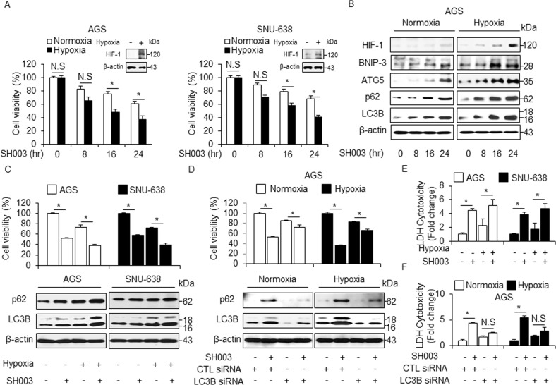
Fig. 7. SH003 induces more autophagic cell death in hypoxia than in normoxia.
a SH003 (400 μg/mL) was added to AGS and SNU-638 cells in a time-dependent manner. Cultures were also exposed to hypoxia or normoxia in a time-dependent manner. Cell viability was assessed using WST-1 assay; *p < 0.05. Western blot analysis was used to validate the targeted changes in HIF-1α expression in hypoxia-induced AGS and SNU-638 cells. b Expression of HIF-1α, BNIP3, ATG5, p62, and LC3B in SH003 (400 μg/mL, 24 h)-treated AGS cells for the indicated times under normoxia or hypoxia. c, e Cell viability analyses, LDH assay, and Western blotting in hypoxia-mediated AGS and SNU-638 cells were analyzed in the presence of SH003 (400 μg/mL, 24 h). β-actin was used as a protein loading control. d, f AGS cells were transfected with control or LC3B siRNA and then exposed to normoxia or hypoxia for 24 h in the presence of SH003 (400 μg/mL, 24 h). Cell viability analyses, LDH assay, and Western blotting were performed for this condition.

