Figure 2:

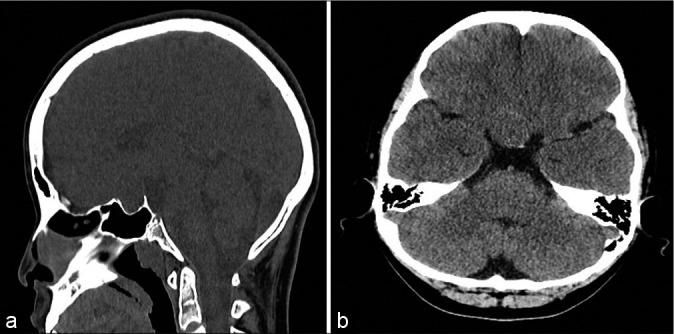
Preoperative imaging noncontrast computed tomography image in sagittal (a) and axial (b) planes shows a sellar mass extending into the suprasellar region. No calcifications are seen in the lesion.

Preoperative imaging noncontrast computed tomography image in sagittal (a) and axial (b) planes shows a sellar mass extending into the suprasellar region. No calcifications are seen in the lesion.