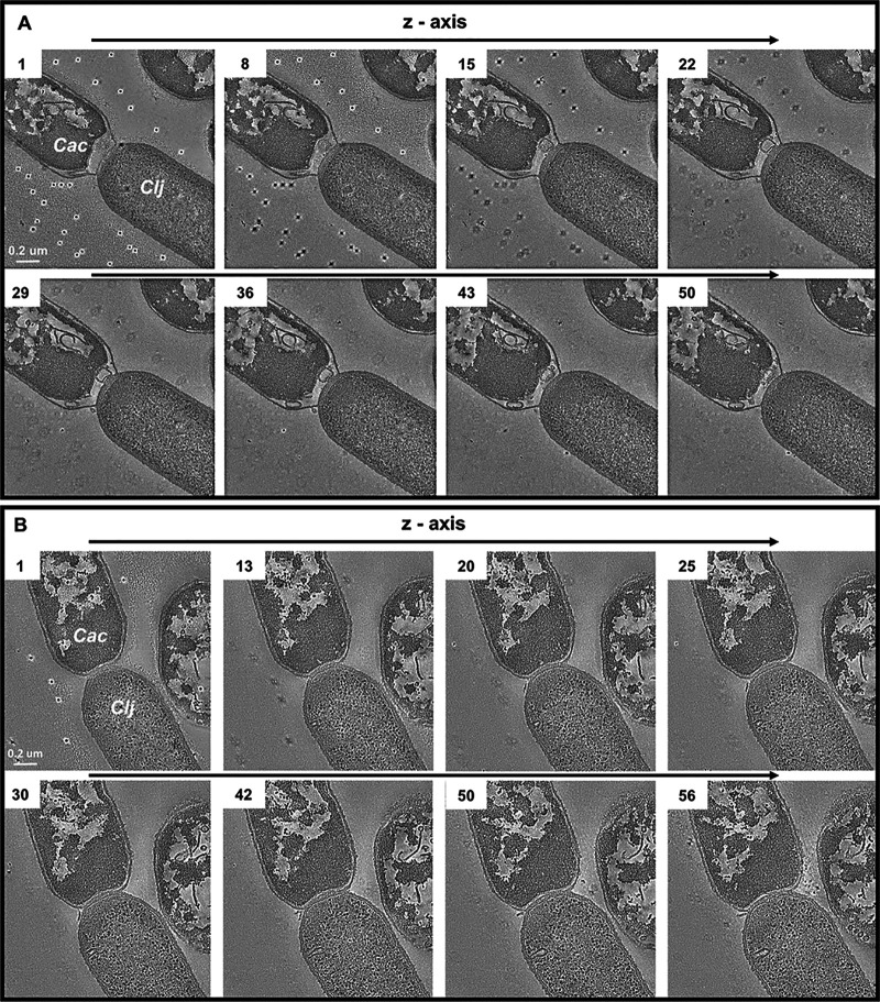
FIG 2.
Electron tomography of C. acetobutylicum and C. ljungdahlii fusion pairs observed in the coculture. (A) TEM tomography: differentiating C. acetobutylicum cells (top left, Cac)) can be easily distinguished from the vegetative homogeneous texture of C. ljungdahlii cells (bottom right, Clj). The cell fusion between the two organisms persists through the entire depth of the tomography (∼150 nm). Dark marks in the background are gold fiducial markers used for aligning the image series. (B) TEM tomography of another C. acetobutylicum-C. ljungdahlii fusion pair.

