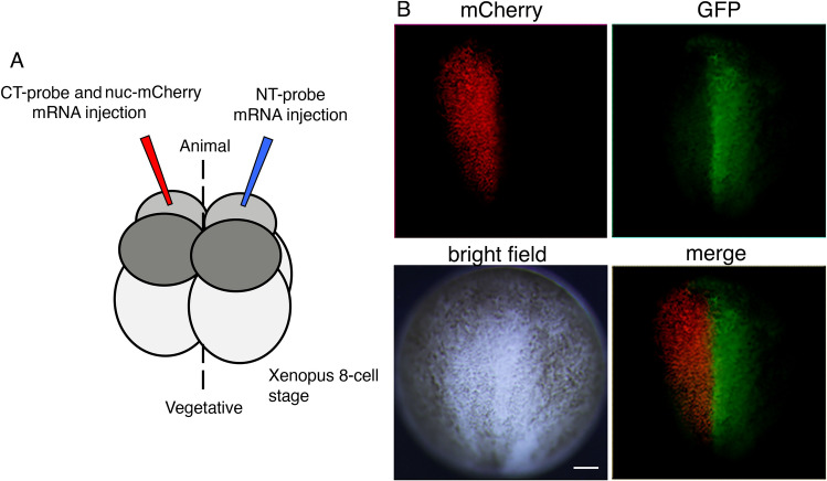
Figure 3.
dGRAPHIC application in Xenopus blastomere. (A) Schema of mRNA injection into 8-cell-stage Xenopus blastomeres. The mRNA of the NT probe is injected in the right animal-smaller blastomere of the Xenopus 8-cell stage, and the mRNAs of the CT probe and nuc-mCherry are injected together into the left animal-smaller blastomere. (B) Embryos are grown to the early neurula stage. Cells expressing the CT probe (red nuclei) are present only within the left half of the neural plate, while GFP signals can be detected only in the right half of the neural plate. Scale bar, 100 μm.

